
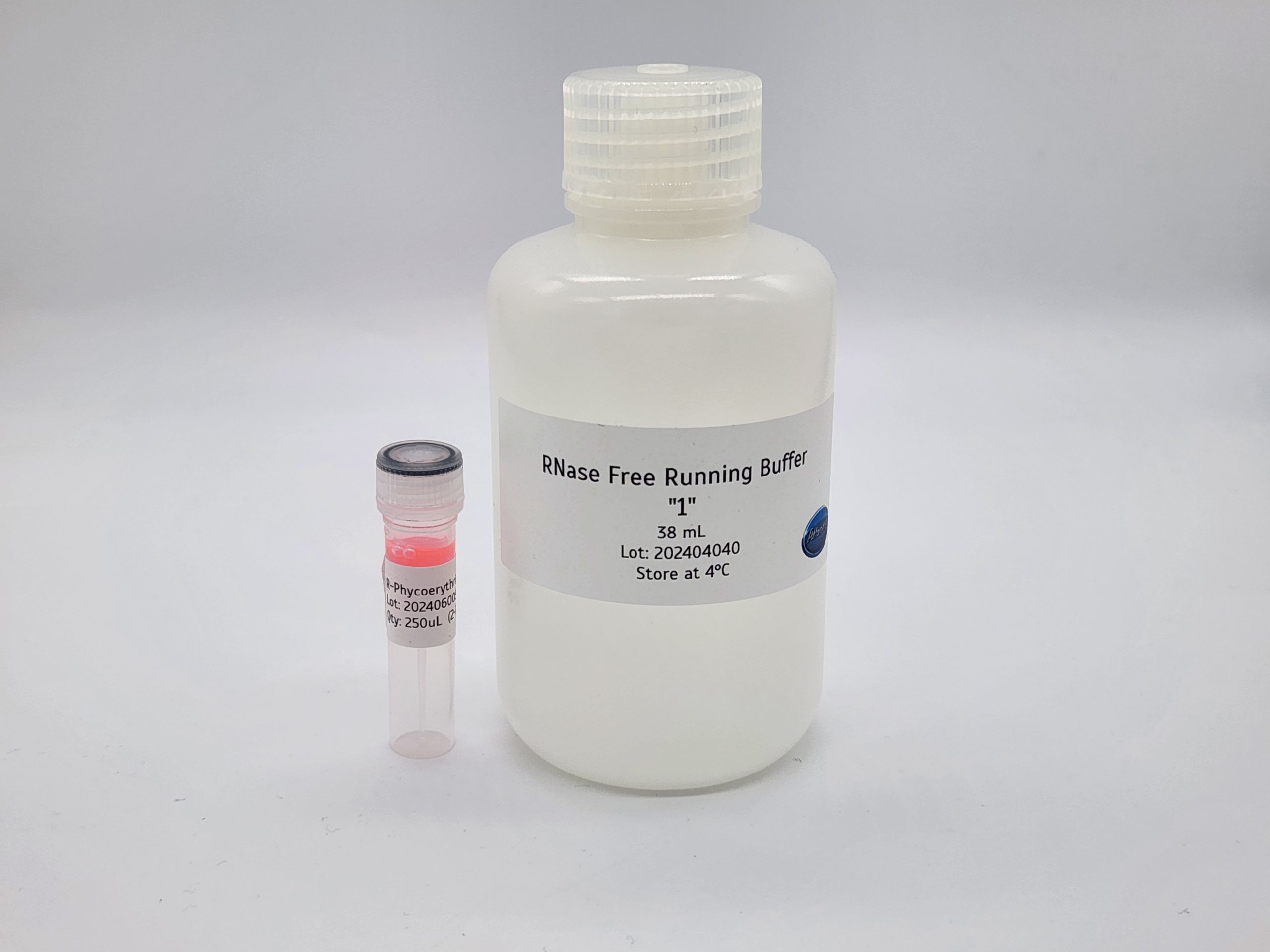


Streptavidin R-Phycoerythrin for Lateral Flow is a ready-to-use streptavidin phycoerythrin conjugate for fluorescent lateral flow assays. R-phycoerythrin (R-PE), a fluorescent protein produced from red algae, is covalently conjugated to streptavidin and supplied as 250 µL of ready-to-use stock together with Attogene RNase-Free Running Buffer #1. The conjugate has an excitation peak of 566 nm and an emission peak of 574 ± 10 nm.
R-PE is a highly absorptive fluorescent molecule with excellent detectability. It is a common choice when a bright signal matters for sensitivity. Because streptavidin binds biotin with very high affinity, the conjugate can label biotin-tagged oligonucleotides and other biotinylated binders in your lateral flow design.
Key Features
- Protein-based fluorescent label: R-phycoerythrin from red algae, designed for lateral flow applications.
- Visible excitation: excitation peak of 566 nm and emission peak of 574 ± 10 nm, per the specification sheet.
- Covalent streptavidin coupling: the phycoerythrin protein is covalently conjugated to streptavidin.
- Consistent lots: continuous culture of the source organisms and high purity support consistent lot-to-lot performance.
- Well-defined protein: very high water solubility and a homogeneous structure with defined molecular weights.
- Running buffer included: supplied with Attogene RNase-Free Running Buffer #1.
Specifications
Each streptavidin phycoerythrin order includes:
- Streptavidin R-PE conjugate: 250 µL of ready-to-use stock
- Running buffer: Attogene RNase-Free Running Buffer #1 (AU2035), 38 mL per the specification sheet
Conjugate specifications, per the specification sheet:
- Fluorophore: R-phycoerythrin protein
- Conjugate: streptavidin, covalently conjugated
- Excitation peak: 566 nm
- Emission peak: 574 ± 10 nm
- Solvent: 50 mM borate buffer, pH 8.3, with 0.05% ProClin 150 and 0.1% BSA
- Storage: 2–8 °C in a dark environment. Do not freeze.
You will also need: a biotin-labeled oligonucleotide or other biotinylated binder, lateral flow strips or another assay format, and a reader or imaging setup that can excite near 566 nm and detect emission near 574 nm.

Who It’s For
This streptavidin phycoerythrin conjugate suits assay developers who want a bright fluorescent label excited by visible light. Typical users include:
- Teams building fluorescent lateral flow assays for nucleic acid or protein targets that carry a biotin tag.
- Developers whose readers or imagers are set up for visible excitation rather than UV.
- Researchers who already use streptavidin gold and want to compare a fluorescent readout on the same assay design.
How to Use Streptavidin Phycoerythrin in Your Assay
The specification sheet gives this starting guide for oligonucleotides:
- Bring the conjugate to room temperature and shake for 30 seconds so it is fully dispersed.
- Add 100–150 µL of RNase-Free Running Buffer #1 to your tube or well.
- Add 0.5–1 µL of the phycoerythrin conjugate. If you dilute it further, use 50 mM boric acid buffer, pH 8.3, with 0.1% BSA and 0.05% ProClin 150.
- Add 0.5–10 pmol of oligonucleotide.
- Run the strip. Run times range from 15 to 45 minutes, depending on the antigen and antibodies used.
To spray or spot the conjugate onto conjugate pads, contact sales@attogene.com for recommendations. Full details are in the streptavidin R-PE specification sheet (PDF).
How the R-Phycoerythrin Is Produced
R-phycoerythrin is a light-harvesting pigment protein found in red algae. The R-PE in this conjugate comes from source organisms grown in continuous culture under fully controlled growth conditions and nutrition. This avoids contamination from extraneous organisms and from waste found in the open ocean.
The proteins are harvested at the optimal stage of the growth cycle for uniform product characteristics. The pigment is then extracted and stabilized within minutes of harvest, which virtually eliminates the risk of protease damage.
For a complete starting point, see the Fluorescent Universal Lateral Flow Assay Kit (R-Phycoerythrin). For a visual readout, compare Streptavidin Colloidal Gold for Lateral Flow. Other fluorescent streptavidin labels include Streptavidin Europium Chelate Microspheres and Streptavidin CdSe/ZnS Quantum Dots.
Storage and Handling
- Store at 2–8 °C away from light. Storage at low temperature increases shelf life and stability.
- Do not freeze. Freezing causes irreversible aggregation and destroys the product.
- Bring to room temperature and shake well before each use.
- Visible particulates or a change in the color or intensity of the solution can mean the conjugate has aggregated. In that case, filter through a 0.45 µm polyvinylidene fluoride (PVDF) filter and keep the filtered product.
Frequently Asked Questions
What are the excitation and emission wavelengths?
The specification sheet lists an excitation peak of 566 nm and an emission peak of 574 ± 10 nm for this streptavidin phycoerythrin conjugate.
What is included with the conjugate?
You receive 250 µL of ready-to-use streptavidin R-PE in 50 mM borate buffer, pH 8.3, with 0.05% ProClin 150 and 0.1% BSA, plus Attogene RNase-Free Running Buffer #1.
How much conjugate should I use per test?
The starting guide uses 0.5–1 µL of streptavidin phycoerythrin with 100–150 µL of running buffer and 0.5–10 pmol of oligonucleotide. Optimize these amounts for your own assay.
Can I freeze the conjugate?
No. Freezing causes irreversible aggregation. Store at 2–8 °C away from light.
Can I spray the conjugate onto conjugate pads?
Contact sales@attogene.com for recommendations on spraying or spotting the conjugate onto conjugate pads.
For research use only. Not for use in diagnostic procedures. Questions? Call 512-333-1330 or email sales@attogene.com.