High quality products to support Pathologists and Biological and Environmental Scientists
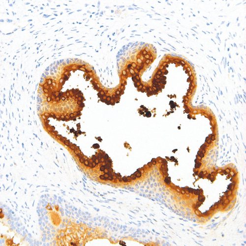
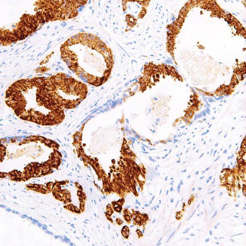
GeneAb™ PSA
$70.00 – $280.00Prostate-Specific Antigen (PSA) is a serine protease of the kallikrein family, that is produced by the prostate epithelium and epithelial lining of the periurethral glands. Although considered prostate-specific, PSA has also been detected in breast tissue, breast tumors, endometrium, adrenal neoplasms, and renal cell carcinomas. Anti-PSA can be used for differentiating high-grade prostate adenocarcinoma from high-grade urothelial carcinoma, as well as for determining the prostatic origin of carcinomas in non-prostate tissues. Anti-PSA recognizes primary and metastatic prostatic neoplasms, but not tumors of nonprostatic origin, and can be useful as an aid to confirm prostatic acinar cell origin in primary and metastatic carcinomas.
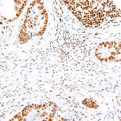
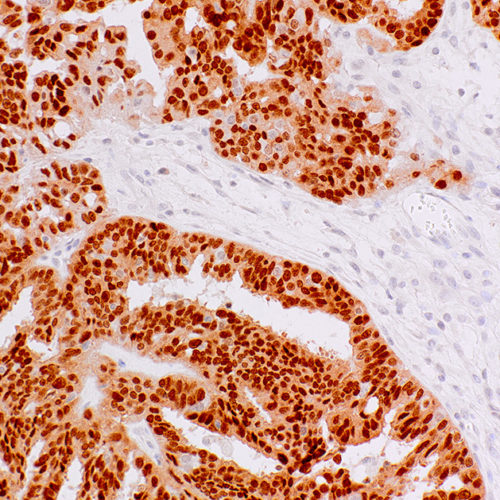
GeneAb™ PMS2
$70.00 – $400.00Postmeiotic Segregation Increased 2 (PMS2) is a DNA repair protein involved in mismatch repair. Mutations and deficiencies in the PMS2 gene have been linked to microsatellite instability, and malignancies such as hereditary nonpolyposis colorectal cancer and endometrial cancer. Expression levels of the PMS2 protein may be useful as a screening tool for Lynch syndrome after a colorectal cancer diagnosis. Anti-PMS2 is recommended to be used as part of a panel along with antibodies against MLH1, MSH2, and MSH6.
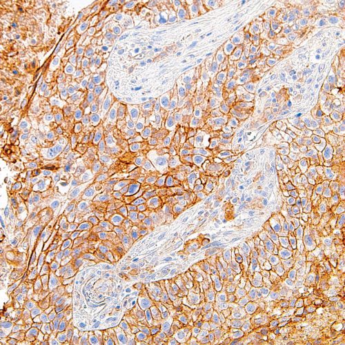
GeneAb™ PD-L1
$145.00 – $625.00Programmed Death-Ligand 1 (PD-L1), also known as CD274 or B7 Homolog 1 (B7-H1), is a transmembrane protein involved in suppressing the immune system and rendering tumor cells resistant to CD8 T cell-mediated lysis through binding of the Programmed Death-1 (PD-1) receptor. Overexpression of PD-L1 may allow cancer cells to evade the actions of the host immune system. In renal cell carcinoma, upregulation of PD-L1 has been linked to increased tumor aggressiveness and risk of death, and, in ovarian cancer, higher expression of this protein has lead to significantly poorer prognosis. PD-L1 has also been linked to systemic lupus erythematosus and cutaneous melanoma. When considered in adjunct with CD8 tumor-infiltrating lymphocyte density, expression levels of PD-L1 may be a useful predictor of multiple cancer types, including stage III non-small cell lung cancer, hormone receptor negative breast cancer, and sentinel lymph node melanoma.
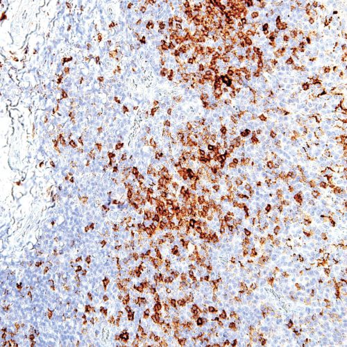
GeneAb™ PD-1
$70.00 – $280.00Programmed Death 1 (PD-1) is a member of the CD28/CTLA-4 family of T-cell regulators, expressed as a co-receptor on the surface of activated T-cells, B-cells, and macrophages. New studies have suggested that the PD-1/PD-L1 signaling pathway may be linked to anti-tumor immunity, as PD-L1 has been shown to induce apoptosis of activated T cells or inhibit activity of cytotoxic T cells. In comparison to CD10 and Bcl-6, PD-1 is expressed by fewer B cells and has therefore been considered a more specific and useful diagnostic marker for angioimmunoblastic T-cell lymphoma. Therapies targeted toward the PD-1 receptor have shown remarkable clinical responses in patients with various types of cancer, including non–small-cell lung cancer, melanoma, and renal-cell cancer.
GeneAb™ PAX-8
$150.00 – $780.00PAX-8 is a member of the paired box (PAX) family of transcription factors, which are key regulators in early development. This protein plays a role in development of thyroid follicular cells and the expression of thyroid-specific genes, with mutations in the PAX-8 gene linked to thyroid follicular carcinomas, atypical thyroid adenomas, and thyroid dysgenesis. The PAX-8 protein is expressed in simple ovarian inclusion cysts and non-ciliated mucosal cells of the fallopian tubes, but is absent from normal ovarian surface epithelial cells. PAX-8 is also not expressed in normal lung or lung carcinomas. Reports have associated PAX-8 expression with renal carcinoma, nephroblastoma, and seminoma, and have indicated PAX-8 as a useful marker for renal epithelial tumors, ovarian cancer, and for differential diagnoses in lung and neck tumors. Anti-PAX-8 can be useful in determining the primary site of invasive micropapillary carcinomas of ovary from bladder, lung, and breast, when used in adjunct with a panel of organ-specific markers such as uroplakin, mammaglobin, and TTF-1.
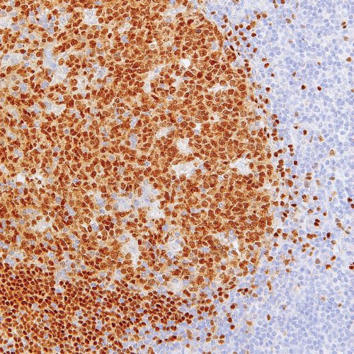
GeneAb™ PAX-5
$50.00 – $255.00PAX-5 is a member of the paired box (PAX) family of transcription factors, which are key regulators in early development. The PAX-5 gene encodes the B-cell lineage specific activator protein (BSAP), whose expression is limited to early stages of B-cell differentiation. Anti-PAX-5 is useful in differentiating between classic Hodgkin’s lymphoma versus multiple myeloma and solitary plasmacytoma, as the protein is expressed in mature and precursor B-cell non-Hodgkin’s lymphomas/leukemias while being absent from the other two conditions. Diffuse large B-cell lymphomas are positive for PAX-5, with the exception of those with terminal B-cell differentiation, and T-cell neoplasms do not stain with Anti-PAX-5.
GeneAb™ p504s
$80.00 – $285.00p504s, also known as α-methylacyl coenzyme A racemase (AMACR), is an enzyme localized in the peroxisome and mitochondria, which functions in β-oxidation of branched chain fatty acids, as well as bile synthesis. AMACR has been clinically indicated as a tissue biomarker for prostate cancer and colorectal cancer, as well as high-grade prostatic intraepithelial neoplasia, a precursor lesion of prostate cancer. p504s overexpression has also been detected in a number of other cancers including ovarian, breast, bladder, lung, and renal cell carcinomas, lymphoma, and melanoma.
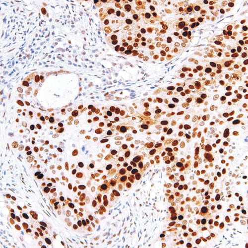
GeneAb™ p53
$70.00 – $280.00p53, also known as tumor protein 53 or TP53, is a tumor suppressor and transcription factor that functions in a number of anti-cancer activities including DNA repair, cell-cycle arrest, and apoptosis in response to DNA damage or other stressors. Mutations in p53 are linked to a number of malignant tumors, including those of the breast, ovarian, bladder, colon, lung, and melanoma. Anti-p53 staining has been used to detect intratubular germ cell neoplasia, and also to distinguish between uterine serous carcinoma and endometrioid carcinoma.
